Freitag, 17. August 2018

In-solution, In-gel, FASP and S-Traping - a voyage through protein sample preparation


When it comes sample preparation in bottom-up proteomics one likes to be as fast, as reproducible and as efficient as possible. Unfortunately, most of the sample preparations are biased towards certain peptide species. In this respect  hydrophobic proteins, such as membrane proteins can be troublesome. Also one should consider sample loss during each preparation step.

However, over the years there have been a couple of techniques established, that are widely used among proteomics researchers. Each of them has advantages and disadvantages.

During In-solution protein digestion protein precipitation in chloroform/methanol is followed by re- solubilization and digestion in 8M urea. This digestion is achieved in reasonable time compared to in-gel digestion but has the disadvantage of introducing sample loss during re-solubilization step.

Second approach is the in-gel preparation, that follows the idea to entrap the protein solution within a polyacrylamid gel matrix (usually after SDS PAGE) and subsequently washing out detergents and performing  protein digestion within the gel. In gel digestion is very time consuming but it is worth though because in most cases you are ending up with a high number of PSMs.

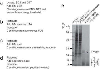
The third technique is called FASP, which stands for filter aided sample preparation and requires about 7h hands on time. FASP tries to combined the advantages of the previously mentioned techniques. In filter aided sample preparation proteins are denatured and kept in solution by SDS. The SDS-protein mixture is subjected onto a filter cartridge, where all proteins are bonded. After an SDS-urea exchange, digestion takes place within the molecular mass cut off filter (be aware of MWCO during selection), releasing peptides whereas undigested proteins remain within the filter and would not contaminate the peptide mixture.

Depending on the geometry of the spin filter and your centrifuge over 50% of the originally used protein amount, ranging from µg to mg, can be recovered on peptide level. I found good SDS PAGE from a nature method paper which served as a control of evaluate the recovery during each step.


A rather new technique is called S-trapping, the S stands for suspension, because the proteins are trapped within a porous network made of quartz (SiO2) while being in suspension. Contaminations and salts have no binding affinity and remain in the flow through. Sample amounts ranging from ng to µg (read somewhere that 250µg is the maximum protein capacity of the silica network)
But it all starts with a common SDS step to solubilize all proteins. Afterwards this SDS micelle is partially broken up and the proteins begin to become partially denatured. This is when the quartz networks kicks in and bonds all of these particulate proteins to prevent them from aggregating with other particulate proteins. Since all the proteins are attached onto the surface of the network proteolytic digestion enzymes have an easy job to access all cleavage sites.

Quartz is a good choice since it provides low metal content (similar to type I silica in HPLC) and low peptide background during digestion. Additionally, one is able to chemically modify the silica surface to perform enrichment of certain peptides after digestion (for example with SDPD, commonly used as crosslinker, for enrichment of cysteine containing peptides, search C-S-trapping).


A recent study comparing all of these 3 sample preparation approaches indicated that S-trapping outperforms in-solution and FASP in terms of identification of unique peptides.


The S-Trapping is commercialized by a company called Protifi. There also provide a unique protease for enhanced b-ion in MSMS fragementation. Great stuff!

Mittwoch, 8. August 2018

In vivo TMT labelling

Tandem Mass Tag (TMT) is a common technique for quantification of peptides at the MS2 level.
The TMT is based on the reaction of the Primary amine at the N-terminus and the lysine with the NHS-ester group of the isotopically labeled tag. Once successfully tagged the peptide mixture is subject to MS analysis were the TMT-peptide displays a label-specific, low mass reporter ion during MS2.

The NHS-based TMT strategy requires all peptides to be freely accessible within a lysate to obtain efficient tagging. TMT can be apply for intact proteins as well, but applying it to intact cell in vivo was new to me.
The authors investigated the labelling efficienicy among different cancer cell lines and stated that in vivo TMT labeling requires an additional enrichment step to achive decent labeling efficiencies which are still lower compared to tagging on the peptide level (roughly 50% of identified peptides were tagged with invivo TMT after enrichment). The enrichment was done using an anti-TMT antibody to pull down all labeled peptides.

Compared to TMT labelling on the peptide level which appeared to be 100% of all identified peptides in vivo labelling with subsequent
Tandem Mass Tag (TMT) is a common technique for quantification of peptides at the MS2 level.
The TMT is based on the reaction of the Primary amine at the N-terminus and the lysine with the NHS-ester group of the isotopically labeled tag. Once successfully tagged the peptide mixture is subject to MS analysis were the TMT-peptide displays a label-specific, low mass reporter ion during MS2.


The NHS-based TMT strategy requires all peptides to be freely accessible within a lysate to obtain efficient tagging. TMT can be apply for intact proteins as well, but applying it to intact cell in vivo was new to me.
The authors investigated the labelling efficienicy among different cancer cell lines and stated that in vivo TMT labeling requires an additional enrichment step to achive decent labeling efficiencies which are still lower compared to tagging on the peptide level (roughly 50% of identified peptides were tagged with invivo TMT after enrichment). The enrichment was done using an anti-TMT antibody to pull down all labeled peptides.
Compared to TMT labelling on the peptide level which appeared to be 100% of all identified peptides in vivo labelling with subsequent immunoprecipation showed rather minor efficiency. However reproducibility was definitely given. Surprisely, the in vivo labelling strategy had no specificity to the location of the proteins. Specifically, a bias towards surface protein has not been revealed.


 showed rather minor efficiency. However reproducibility was definitely given. Surprisely, the in vivo labelling strategy had no specificity to the location of the proteins. Specifically, a bias towards surface protein has not been revealed.


Freitag, 3. August 2018

New features in skyline - LC-IMS-CID-MS and contaminations library


There is a lot going on in the skyline world lately. Originally, open source software skyline from the MacCoss Lab which started out as analysis tool for label-free quantification and MRM analysis of MS data.
Over years a lot of features have been added because more comprehensive analysis was requested by the users or new instruments using new scan modes have been introduced to the market. That's why recently ion mobility functionality has been added, including the entire LC-IMS-CID-MS workflow.


However, yet another skyline feature caught my attention in the current JASMS. A contamination library has been integrated into skyline. It contains over 684 common MS contaminations, which can be used as an transition list for MS1 filtering and is also provides a approach to determine unknown contaminations via a mass-to-formula tool. The mentioned transition list can be downloaded in the public repository panorama.


CID and CIU

CID stands for collision-induced-dissociation a common fragmentation technique to create tandem MS spectra (MS2). In CID precursor ions are accelerated and subsequently injected into a ion guide filled with inert neutral gas molecules, typically N2, where the analyte of interest undergoes single or multiple collisions. The charge of the analyte, the accelerating (collisional) voltage and the gas pressure can determine the extent of collisional Impact.

Ions are vibrational excited by the collision(s) with a time frame of a few femtoseconds. Depending on the chemical bonds present the ions can break apart into charged, radical or neutral fragments. In proteomics weaker bonds, such a post-translational modification, tend to get lost during CID.
In beam type instruments a special version of CID, In Source CID, can facilitate MS3 fragmentation for deeper structural elucidation. Herein, ion guides on the front end of the MS serve as a 2nd collision device for fragmentation of all ion species injected. Out of this very complex MS2 spectrum precursors can be selected for an MS3 in the actual collision cell downstream.  

When it comes to analysis of intact proteins and protein complexes having high molecular weights, In-Source CID at elevated pressure, can be applied but in most cases it directly leads to dissociation. Secondary or high order protein structures cannot be detected easily.
Besides the dissociative nature of CID for large biomolecules there is a much elegant appoarch that utilizes collisional activation to study structures intactly. It is called CIU, which stands for collisional induced unfolding, and can be conducted with an ion mobility detector coupled to an MS.
During a CIU experiment intact biomolecules, such as antibodies, are ionzied and undergo a stepwise increase of collision heating which induces a gradual change in conformation (unfolding).
For every stepped potential an ion mobility scan, plus the nested MS scans, are recorded so that one can follow the structural changes. These multidimensional datasets (see image) help to distinguish isoforms, determine number for disulfide bonds and degree of modifications or monitor ligand binding.

Source: https://www.sciencedirect.com/science/article/pii/S1367593117301266?via%3Dihub#fig0010